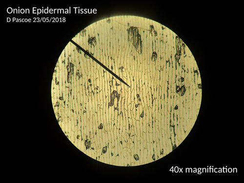

Here are some photos of Onion Epidermal cells and Human Epithelial cells (cheek cells) at 40x, 100x and 400x magnification. Also included are photos of a 1/0.01mm micrometer slide so students can measure the size of the individual cells.
Something went wrong, please try again later.
I was delighted to find this but it would be improved by 1) the 'graticule' slides are actually photos of the micrometer slide at different magnifications, as is evidenced by the fact that it gets larger as the magnification increases. The graticule is ANOTHER item which stays in the eyepiece so the size of its scale does not change. The way the two work together is that the graticule is placed in the eyepiece and used as an arbitrary scale to determine at each power how long each graticule unit is, by comparing it with the micrometer slide. 2) It would be absolutely wonderful if you could do more micrometer slide photos with the graticule in place so students could do this calculation. 3) If you could then have a selection of photos of various microscope slides with only the graticule in place so students could calculate thew length/width of cell parts that would also be very gratefully received!
great resource thank you
Report this resourceto let us know if it violates our terms and conditions.
Our customer service team will review your report and will be in touch.
£0.00