pptx, 3.01 MB
pptx, 3.01 MB

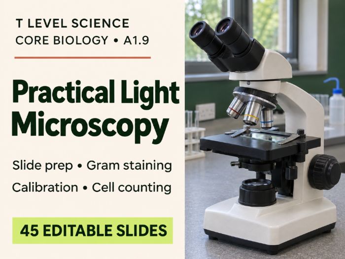
Editable T Level Science A1.9 PowerPoint on light microscopy: slide preparation, Gram staining, graticule calibration and haemocytometer cell counting.

This 45-slide PowerPoint covers A1.9: Using a Light Microscope for Level 3 T Level Science Core Biology. Students progress from preparing and focusing a slide to measuring specimens and calculating cell concentration. The deck is organised into three teaching sections, with suggested pause points and teacher notes to help you plan around equipment access.

What students will learn

  • Microscope operation: identify key controls, calculate total magnification, focus at low power and troubleshoot common problems when changing objectives.
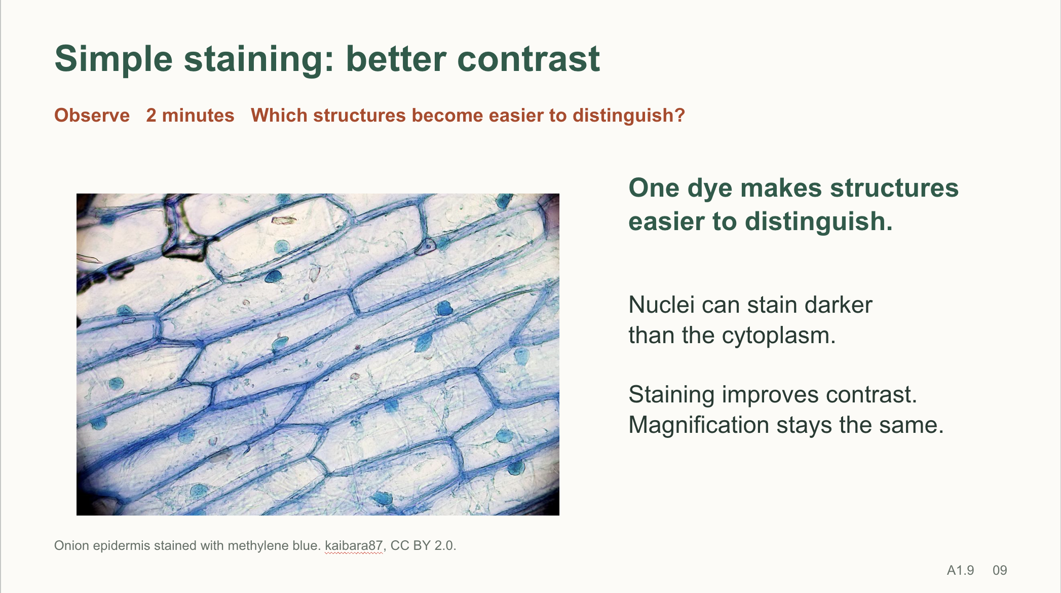
  • Slide preparation and staining: prepare a stained onion wet mount, explain how simple stains improve contrast and compare simple staining with differential staining.
  • Gram staining: explain the purpose of each reagent, interpret Gram-positive and Gram-negative results from a real micrograph and use controls to diagnose staining errors.
  • Calibration and measurement: use a stage micrometre to calibrate an eyepiece graticule, calculate actual specimen size in micrometres and explain why each objective needs its own calibration.
  • Haemocytometer counting: apply a consistent boundary rule, explain where the ×10⁴ conversion factor comes from for the stated chamber volume, correct for dilution and distinguish cell concentration from total cell number.

What’s included

  • Real microscopy photographs and a labelled counting chamber illustration.
  • Timed pair tasks, troubleshooting scenarios and an optional three-station practical circuit covering preparation and focus, calibration and measurement, and yeast cell counting.
  • Worked calculations, practice data and selected click-to-reveal answers so students can attempt each task before seeing the method.
  • An original six-mark practice question with marking points, plus an exit ticket and answers.
  • Teacher notes, source credits and classroom alternatives using supplied images and data when specialist equipment is limited.

Use the editable PowerPoint for practical preparation, classroom teaching or revision. Subtle transitions and selected animations let you control the pace of demonstrations, worked examples and feedback.

Get this resource as part of a bundle and save up to 20%

A bundle is a package of resources grouped together to teach a particular topic, or a series of lessons, in one place.

Bundle

T Level Science - Biology A1: Cells & Tissues Bundle

T Level Biology A1.1 to A1.9: nine editable PowerPoints on cell structure, stem cells, magnification, resolution and practical microscopy. Teach **A1: Cells and Tissues** from the Pearson T Level Technical Qualification in Science with this complete set of nine Core Biology PowerPoints. The lessons take students from comparing cell structures to choosing a suitable microscope, interpreting images and carrying out practical measurements. Teach the decks in sequence or use individual topics for targeted revision. **Nine PowerPoints included** - **A1.1 Prokaryotic and eukaryotic cells:** compare the structure and ultrastructure of the two cell types and use evidence to classify unfamiliar examples. - **A1.2 Eukaryotic cell ultrastructure:** identify cellular components and explain how their structures support their functions. - **A1.3 Plant and animal cells:** compare shared and distinctive structures and relate them to what the cells do. - **A1.4 Cell specialisation and stem cells:** explore differentiation, stem cell sources and potency, and adaptations in nine specialised cell types. - **A1.5 Prokaryotic cell ultrastructure:** examine the structures of a typical bacterial cell and explain their roles. - **A1.6 Magnification and resolution:** distinguish a larger image from one that reveals more detail. - **A1.7 Magnification calculations:** practise unit conversions, rearrange the magnification equation and use scale bars to calculate actual size. - **A1.8 Microscopy principles and limitations:** compare microscopy techniques and justify which one suits a particular investigation. - **A1.9 Practical light microscopy:** prepare and focus slides, interpret Gram stains, calibrate an eyepiece graticule and estimate cell concentration using a haemocytometer. **What teachers get** Across the bundle, you’ll find diagrams and real micrographs, timed activities with clear instructions, worked calculations, and original practice questions with answers or suggested marking points. Teacher notes support delivery, and selected slides reveal steps or answers on click so students can attempt the task first. The practical microscopy deck also includes a three-station circuit and alternatives using supplied images and data when specialist equipment is limited. All nine PowerPoints are editable, so you can adapt the pace, activities and examples for your Level 3 group.

£20.59

Reviews

Something went wrong, please try again later.

This resource hasn't been reviewed yet

To ensure quality for our reviews, only customers who have purchased this resource can review it

Report this resourceto let us know if it violates our terms and conditions.
Our customer service team will review your report and will be in touch.